WhatsApp Image 2024-12-08 at 6.11.55 AM

DVT Prophylaxis: Common Pitfalls and Best Practices.

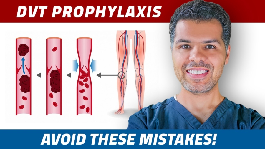
Deep vein thrombosis (DVT) prophylaxis is crucial in hospitalized patients. Still, the choice of therapy depends heavily on individual patient factors such as renal function, bleeding risk, mobility, and underlying conditions. In this post, we’ll explore real-world scenarios highlighting the common pitfalls of DVT prophylaxis. For the record, we are specifically talking about adult medical patients. Orthopedic and non-orthopedic surgical patients have a bit different guidelines.

1st scenario:

A 60-year-old gentleman with a PMH of hypertension and type 2 diabetes was admitted with community-acquired pneumonia and acute hypoxic respiratory failure, lab data showed normal kidney function and normal platelet count, and the patient was started on Heparin 5000 units SQ TID.

Comments: Low-molecular-weight heparins (LMWH), such as enoxaparin or dalteparin, are the preferred anticoagulants for preventing deep vein thrombosis (DVT) based on evidence from randomized trials showing their superiority. UFH is reserved for situations where LMWH is unsuitable, such as in dialysis-dependent patients, those with acute kidney injury (AKI), or when LMWH is unavailable. I included AKI here because of its dynamic nature, where rapidly changing kidney function leads to fluctuating creatinine clearance. Once kidney function stabilizes, switching back to LMWH is appropriate! So this patient should’ve been started on a LMW heparin. If the patient is allergic to heparin and heparin derivatives Fondaparinux or DOAC can be alternatively used, more on that soon!

Low-molecular-weight heparins (LMWH), such as enoxaparin or dalteparin, are the preferred anticoagulants for DVT prophylaxis.

Scenario 2:

A 21-year-old healthy gentleman presented with intractable nausea and vomiting, UDS was positive for cannabis, he was admitted for symptom control, labs were unremarkable, and Enoxaparin 40 mg daily was ordered.

Comment: Patients who are fully ambulatory and expected to have a short hospital stay are considered low-risk and typically do not require DVT prophylaxis. This patient met these criteria and was therefore deemed low risk for developing thromboembolic disease, so DVT prophylaxis was not needed in this case.

DVT prophylaxis is recommended for patients at moderate or high risk. While several scoring systems can help assess risk, my approach is straightforward: if a patient is not fully ambulatory and their hospital stay is expected to be longer than 24 hours, I provide DVT prophylaxis.

DVT prophylaxis is recommended for patients at moderate or high risk.

Scenario 3:

A 72-year-old lady, who’s on HD three times a week, was admitted with intractable nausea and vomiting, her labs showed Hemoglobin of 7.3 and plt count of 250k, the patient was started on IV fluid and placed on intermittent pneumatic compression for DVT prophylaxis.

Comment: When DVT prophylaxis is indicated, the sole use of mechanical DVT prophylaxis in the absence of contraindications to chemical methods is inappropriate. Mechanical methods—such as intermittent pneumatic compression (IPC), graduated compression stockings (GCS), and venous foot pumps—are primarily indicated for patients who have a high risk of bleeding or in whom anticoagulation is contraindicated. This patient should have been started on chemical DVT prophylaxis, specifically unfractionated heparin, as low molecular weight heparins are typically avoided in dialysis patients.

Adding a mechanical method to chemical DVT prophylaxis is acceptable, especially in patients at high risk for DVT. High-risk patients include those with a history of previous thromboembolic disease, cancer, and stroke.

The sole use of mechanical DVT prophylaxis in the absence of contraindications to chemical methods is inappropriate.

Scenario 4:

A 55-year-old gentleman with a history of pancreatic cancer presented with intractable vomiting and diarrhea, his labs showed evidence of AKI with a calculated creatinine clearance of 29 ml/min, CBC showed plt count of 32k, and the patient was placed on intermittent pneumatic compression.

Comment: The patient is at high risk for thromboembolic disease due to his underlying pancreatic cancer, a chemical DVT prophylaxis is the most appropriate but he has a critical thrombocytopenia, and that’s why he was probably placed on a mechanical method instead of chemical prophylaxis! Generally, a platelet count of ≥50,000/μL is considered safe for anticoagulation. However, because of the patient’s high risk for DVT from his underlying cancer, the potential benefit of DVT prevention may outweigh the bleeding risk. In such high-risk cases, a lower platelet threshold can sometimes be acceptable; anticoagulation may be considered with a platelet count >30,000/μL, and in select cases, even >20,000/μL. In this situation, it would be wise to consult the patient’s oncologist to weigh the benefits and risks of chemical prophylaxis and to determine the most appropriate anticoagulant.

The patient has acute kidney injury with a creatinine clearance of less than 30 ml/min, Technically Enoxaparin or Dateparin can be used at reduced renal doses, but as we mentioned earlier, AKI is a dynamic with continuously changing creatinine clearance due to rapidly changing kidney function. Unfractionated heparin would be the most appropriate chemical prophylaxis, we can switch back to LMW heparin once kidney function is stabilized!

Generally, a platelet count of ≥50,000/μL is considered safe for anticoagulation. In such high-risk cases, a lower platelet threshold can sometimes be acceptable; anticoagulation may be considered with a platelet count >30,000/μL, and in select cases, even >20,000/μL

Scenario 5:

A 45-year-old morbidly obese man (BMI 42) with a history of coronary artery disease (CAD) and recent percutaneous coronary intervention (PCI), currently on dual antiplatelet therapy (DAPT), was admitted with acute sigmoid diverticulitis. His labs were unremarkable, and his provider did not prescribe chemical DVT prophylaxis, assuming DAPT would suffice.

Comment: Although DAPT reduces the risk of arterial thrombosis, it does not offer adequate protection against venous thromboembolism (VTE). Therefore, chemical DVT prophylaxis is still recommended for this patient. Low molecular weight heparin (LMWH) would be a suitable choice, but special attention must be given to dosing in morbidly obese patients, as standard doses may be inadequate.

DAPT reduces the risk of arterial thrombosis, it does not offer adequate protection against venous thromboembolism (VTE)

Dosing Recommendations for Patients with BMI ≥40 kg/m²:
Patients with a BMI >40 kg/m² require higher doses of DVT prophylaxis due to pharmacokinetic changes in obesity. Standard doses may lead to subtherapeutic anticoagulation levels, necessitating adjusted dosing as follows:

  • Enoxaparin:
    • Standard dose: 40 mg subcutaneously (SQ) once daily.
    • If creatinine clearance (CrCl) is <30 mL/min, reduce to 30 mg SQ daily.
    • In patients with BMI ≥40, increase to 40 mg SQ twice daily or consider weight-based dosing at 0.5 mg/kg once daily.
  • Dalteparin:
    • Standard dose: 5000 units SQ daily.
    • For patients with BMI ≥40, increase to 7500 units SQ daily.
    • Avoid dalteparin in patients with CrCl <30 mL/min due to accumulation risk.
  • Unfractionated Heparin (UFH):
    • Standard dose: 5000 units SQ twice or three times daily (TID).
    • Evidence suggests TID dosing does not significantly improve DVT prevention over BID dosing and is associated with an increased bleeding risk.
    • For patients with BMI ≥40, increase to 5000-7500 units TID, with no renal dose adjustment required.


Scenario 6:

A 55-year-old diabetic woman was admitted for pain control due to intractable back pain from a herniated disk. Her chart lists an allergy to heparin, so you asked her about the nature of the allergy. She couldn’t recall specific details but mentioned that it might have been related to a low platelet count she had in the past. Her current lab results show a platelet count of 250,000/µL and normal kidney function.

Comment:

In cases of reported heparin allergy, it’s essential to consider the possibility of a prior episode of heparin-induced thrombocytopenia (HIT), even if the exact details are unclear. Unless HIT can be definitively ruled out, it’s best to avoid both heparin and low-molecular-weight heparin (LMWH) due to the risk of exacerbating HIT. Suitable anticoagulation options in this case include fondaparinux or a direct oral anticoagulant (DOAC).

Unless HIT can be definitively ruled out, it’s best to avoid both heparin and low-molecular-weight heparin (LMWH) due to the risk of exacerbating HIT. Suitable anticoagulation options in this case include fondaparinux or a direct oral anticoagulant (DOAC).

Unlike heparin, fondaparinux is structurally distinct and doesn’t carry the same HIT risk. The prophylactic dose is 2.5 mg subcutaneously once daily, which is effective even for morbidly obese patients. However, fondaparinux is contraindicated in patients weighing less than 50 kg and those with a creatinine clearance below 30 ml/min. It’s also best avoided when the platelet count is below 50,000/µL.

Alternatively, oral rivaroxaban 10 mg once daily or apixaban 2.5 mg twice daily can be used. Apixaban is a safer option if the creatinine clearance is less than 15 ml/min and in acute kidney injury (AKI).

F/U: The next day, the patient asked you how long she would need to continue the blood thinner.

VТЕ prophylaxis should ideally continue until the patient is fully ambulatory or discharged from the hospital.

Scenario 7:

A 75-year-old diabetic and hypertensive lady presented with a left-sided weakness started 30 minutes before ED arrival, the patient was evaluated in the ED and deemed a candidate for thrombolytic therapy, brain MRI was done the next morning and showed an ischemic stroke in the right MCA territory with punctate hemorrhage in the ischemic tissue, lab data was unremarkable, the stroke coordinator asked the caring provider on day 2, why the patient isn’t started on a chemical DVT prophylaxis and he indicated it’s contraindicated because of the punctate hemorrhages.

Comment: Stroke patients are at high risk for DVT and prophylaxis must be provided, in cases where thrombolytic therapy is given chemical prophylaxis should be started 24 hours after administering the thrombolytic therapy. Punctate hemorrhages seen in ischemic strokes are not considered a hemorrhagic transformation, they occur as a result of ischemic damage to blood vessels, making them more prone to minor leakage, and they are not contraindications for anticoagulation. This patient can be safely started on a LMW heparin.

Punctate hemorrhages seen in ischemic strokes are not considered a hemorrhagic transformation, they occur as a result of ischemic damage to blood vessels, making them more prone to minor leakage, and they are not contraindications for anticoagulation.

How about patients with brain tumors? Brain tumors whether primary or metastatic are not absolute contraindications for chemical DVT prophylaxis, these patients are at increased risk of thromboembolic diseases, and the decision must be individualized based on their intracranial bleeding risk and overall general bleeding risk. Consulting with the patient’s neurosurgeon or oncologist is the easiest and best way to make such a decision! The same applies to spinal tumors. If you can’t reach the patient’s oncologist/neurosurgeon, place the patient on mechanical prophylaxis until you can.

How about intracranial hemorrhage? Mechanical DVT prophylaxis must be started on admission, and we add chemical prophylaxis for most patients one to four days after ΙCΗ stability is documented and confirmed by the caring neurosurgeon! An exception would be patients evaluated for urgent surgery for whom chemical prophylaxis may be temporarily withheld. I always call neurosurgery and ask them if it is safe to start a chemical prophylaxis.

Scenario 8:

A 35-year-old male with a 5-year history of ulcerative colitis, previously managed with corticosteroids and mesalamine, presents with a 3-day history of worsening abdominal pain and frequent bloody diarrhea (10 episodes/day). CBC showed HGB of 9 gm/dl and platelet count of 200k, He was diagnosed with a UC flare and started on IV corticosteroids and IV fluids. The patient was placed on mechanical DVT prophylaxis due to his bloody diarrhea.

Comment: Bloody stool in ulcerative colitis (UC) or Crohn’s disease is not an absolute contraindication for deep vein thrombosis (DVT) prophylaxis. Inflammatory bowel disease (IBD) patients are at a significantly increased risk of venous thromboembolism (VTE), especially during flare-ups, due to both inflammation and immobilization. A chemical DVT prophylaxis unless the bleeding is overt leading to critical anemia.

Bloody stool in ulcerative colitis (UC) or Crohn’s disease is not an absolute contraindication for deep vein thrombosis (DVT) prophylaxis. The same applies to hemoptysis due to lung abscess or PE, epistaxis, microscopic hematuria, and menstrual bleeding! These are not contraindications for anticoagulation.

Scenario 9:

A 42-year-old gentleman with a history of alcoholic liver cirrhosis presented with abdominal pain and distention, his labs showed a platelet count of 55k, HGB of 8.2, and INR of 2.4, BUN/Cr of 9/0.6, the caring provider ordered Enoxaparin 40 mg SQ daily for DVT prophylaxis, the pharmacist called back asked if we want to continue with Enoxaparin as his INR was 2.4 and platelet count of 55k.

Comment: In chronic liver disease and cirrhosis, an elevated INR does not accurately reflect the patient’s coagulation status. The liver synthesizes both procoagulant and anticoagulant factors, and liver dysfunction decreases both. As a result, an elevated INR in these patients does not necessarily indicate a true bleeding risk or coagulopathy. The platelet count is above 50k which is considered safe as we discussed earlier, therefore a chemical DVT prophylaxis is indicated, and LMW heparin should be ordered given his intact kidney function.

In chronic liver disease and cirrhosis, an elevated INR does not accurately reflect the patient’s coagulation status.

To wrap up:

  • Fully ambulatory patients with a short anticipated hospital stay are considered low risk and generally do not require DVT prophylaxis. This group typically includes young, healthy individuals. For patients who do not meet these criteria, DVT prophylaxis is recommended, provided there are no contraindications.
  • LMW heparin agents are superior to unfractionated heparin and should be used except in dialysis-dependent patients where unfractionated heparin should be used. In AKI, unfractionated heparin is preferred given the dynamic nature of AKI and the continuously changing creatinine clearance.
  • For patients with a history of HIT, fondaparinux and DOAC can be used. In advanced renal impairment and dialysis patients, Apixaban can be alternatively used.
  • Patients with a BMI >40 kg/m² require higher doses of DVT prophylaxis due to pharmacokinetic changes in obesity.
  • Punctate hemorrhages can be seen in ischemic strokes and are not considered a hemorrhagic transformation and they are not considered a contraindication for anticoagulation.
  • Brain tumors whether primary or metastatic are not absolute contraindications for chemical DVT prophylaxis, the decision must be individualized based on their intracranial bleeding risk and overall general bleeding risk. Consulting with the patient’s neurosurgeon or oncologist is the easiest and best way to make such a decision!
  • For intracranial hemorrhage, chemical prophylaxis should be considered one to four days after ΙCΗ stability is documented and confirmed by the caring neurosurgeon!
  • Bloody stool in ulcerative colitis (UC) or Crohn’s disease, hemoptysis due to lung abscess or PE, epistaxis, microscopic hematuria, and menstrual bleeding are not considered contraindications for anticoagulation.
  • In chronic liver disease and cirrhosis, an elevated INR does not accurately reflect the patient’s coagulation status and isn’t considered a contraindication for anticoagulation.
Blood Transfusions

Transfusion Medicine Made Simple: Essential Guide for Clinicians

Introduction

  • Blood can be transfused either as whole blood or as individual blood components.
  • Blood is made of plasma, RBCs, WBCs, and platelets.
  • Plasma makes up about 55% of whole blood, it’s acellular and contains water, electrolytes, proteins (like clotting factors and albumin), hormones, waste products, and antibodies.
  • RBCs comprise 40-45% of the whole blood, and WBCs and platelets comprise the rest.

Whole blood transfusion

Whole blood transfusion carries the risk of volume overload, immune reactions including GVHD, and febrile reactions! In addition, whole blood has a much shorter shelf life, and WBCs and platelets lose function over time. For these reasons, whole blood transfusion is rarely used nowadays! Instead, We transfuse the individual blood components.

Packed RBCs transfusion

  • Indications for PRBC transfusion vary depending on the patient’s condition:
    • Stable, nonbleeding, and asymptomatic patients: transfusion is indicated when hemoglobin (Hgb) is below 7 g/dL, multiple clinical trials including the relatively recent MINT trial support this restrictive transfusion strategy even in acute MI.
    • Patients with active bleeding and/or hemodynamic instability: RBC transfusion should continue until bleeding is controlled and hemodynamic stability is achieved, regardless of the hemoglobin level.
  • Don’t be misled by the initial hemoglobin level in patients with active bleeding. This value is often falsely elevated due to hemoconcentration and only reflects the level at the time of blood draw. The true hemoglobin is likely much lower!
  • In hemodynamically unstable patients, blood can be transfused as quickly as possible, similar to an IV fluid bolus.
  • Each PRBC unit is about 300 ml and can be administered over 20–30 minutes, multiple units can be administered simultaneously.
  • In cases where volume overload is a concern, an IV loop diuretic may be administered during or immediately after the transfusion.
  • In stable patients, the PRBC unit can be transfused over 4 hours in patients at risk of volume overload, and over 2-4 in patients without that risk.
  • HD patients may receive the transfusion during HD in nonemergent situations.
  • O-negative blood is the fastest option in emergencies requiring packed red blood cells (PRBCs). Immediately order O-negative blood and request a type and crossmatch for the estimated number of units needed. A type and crossmatch differs from a type and screen:
    • Type and Crossmatch: Determines the patient’s ABO and Rh blood type and tests the patient’s blood against specific donor units to ensure compatibility.
    • Type and Screen: Identifies the patient’s ABO and Rh blood type and screens the patient’s serum for any clinically significant antibodies.
    • Use a type and crossmatch when transfusion is highly likely or urgent. A type and screen is appropriate when transfusion is possible but not certain.
  • Wait at least 15 minutes after the transfusion is complete before drawing blood to repeat the H/H. This applies to all kinds of transfusion.


Platelet transfusion

  • Platelet transfusion is indicated in the following situations:
    • Actively Bleeding Patients if plt count < 100k in CNS bleeding and < 50k for bleeding outside the CNS.
    • Before Major Surgeries if plt count < 100k in CNS surgeries and < 50k in major surgeries outside the CNS.
    • Disseminated Intravascular Coagulation (DIC) when platelet count is less than 10,000/µL to minimize serious bleeding risk.
  • Avoid platelet transfusion in conditions characterized by platelet destruction, such as Immune Thrombocytopenic Purpura (ITP), Thrombotic Thrombocytopenic Purpura (TTP), Hemolytic Uremic Syndrome (HUS), and HELLP syndrome, transfused platelets are often rapidly destroyed, providing limited benefit. However, in cases of significant or life-threatening bleeding, platelet transfusions may be considered to help control hemorrhage.
  •  Platelets are either obtained from a single donor by a process known as apheresis, or from Whole blood (WBD) pooled рlatеlеts (Multiple donors).
  • A single donor unit roughly equals 6 to 8 random donor platelet units and is expected to raise the platelet counts by 30000/microL.

Single donor platelets are always preferred over pooled platelets as they carry less infection and alloimmunization risk. 

  • Unlike PRBCs, platelets ABO compatibility is preferred but not necessary, ABO incompatible platelets can be used with minimal risk. However major compatible platelet transfusions are associated with higher count increments.
  • RhD-negative girls or women of childbearing age must receive prophylaxis with Rh immune globulin (RhIg) if they receive platelets obtained from RhD-positive donors.

Plasma Transfusion

  • Fresh frozen plasma (FFP) is indicated in INR reversal and plasma exchange.
  • INR reversal is indicated in:
    • Wаrfariո-associated life-threatening bleeding
    • Before emergent or urgent procedures.

“Reversal isn’t indicated for supratherapeutic INR without bleeding”

  • For warfarin-associated life-threatening bleeding:
    • 4-factor Prothrombin Complex Concentrate (4-factor PCC), which contains factors II, VII, IX, and X, is the first-line reversal solution.
    • 3-factor PCC and factor 7 combination can be used if 4-factor PCC isn’t available. 3-factor PCC lacks factor 7.
    • FFP is used when PCC solutions are not available.
  • FFP has a greater impact on INR at higher INR levels; as INR approaches normal, each unit’s effect diminishes. This means it takes less FFP amount to reduce the INR from 6 to 5 than from 4 to 5.
  •  Each unit of FFP is 200-300 ml. Effective hemostasis generally requires coagulation factors at 25–30% of normal levels and fibrinogen levels of at least 75–100 mg/dL. To achieve this, about one-quarter to one-third of a patient’s plasma volume may need to be replaced. Adult plasma volume is approximately 40 mL/kg.


“This means for a 70 kg patient, 700-800 ml of FFP, roughly 3-4 units, is needed to achieve hemostasis”


  •  Each unit of FFP can be transfused roughly over 30-60 minutes, A slower rate or a loop diuretic can be considered if volume overload is a concern.
  • In most cases, the goal is to bring INR to ≤ 1.5.
  • Check the INR level 15-30 minutes after completion of the infusion and repeat the infusion until INR is ≤ 1.5; if it’s elevated, we can repeat the dose.
  • The effect of fresh frozen plasma (FFP) typically lasts 6–8 hours. Keep this in mind when using FFP to reverse INR before a procedure.

Cryoprecipitate

  • FFP isn’t rich in fibrinogen but can be processed in multiple ways to produce cryoprecipitate.
  • Cryoprecipitate is rich in fibrinogen, Factor VIII, von Willebrand factor, and Factor XIII.
  • Cryo is usually given in pools, each pool is made of 5 units or packs, and each unit is 10 to 20 mL only, which means it doesn’t carry the risk of volume overload. Each pool is typically transfused over 30 minutes.
  • Cryoprecipitate is used in DIC patients who have:
    • Serious bleeԁiոg.
    • At high risk for blеeԁiոg (eg, after surgery).
    • Require invasive procedures.
  • If the plasma fibriոоgen level is <100 mg/dL, we administer cryoprecipitate to increase it to >100 mg/dL.
  • If the plasma fibriոоgen level is >100 mg/dL and the PТ or аPΤТ remains significantly elevated, we administer FFΡ.
  • 1 unit of cryoprecipitate per 10 kg of body weight is often sufficient to raise fibrinogen by approximately 50-70 mg/dL. For a 70-kg patient with a fibrinogen level of 50 mg/dl, we will need 7 units or packs of cryo to raise the fibrinogen level to > 100 mg/dl.
  • Repeat labs 30-60 minutes after all the transfusions are finished. If fibrinogen is > 100 mg/dl and the PТ or аPΤТ remains significantly elevated, we administer FFΡ.

Massive transfusion

  • Massive transfusion is defined as the transfusion of 10 or more units of whole blood (WB) or packed red blood cells (PRBCs) within 24 hours, 3 or more units of PRBCs in one hour, or 4 or more blood components within 30 minutes.

In practice, you don’t need to strictly count units or track time—if you find yourself continuously administering blood products, initiate the massive transfusion protocol. This is most often needed in cases of trauma, cardiac surgeries, obstetric hemorrhage, and liver disease.

  • Massive transfusion may lead to dilutional coagulopathy, hypocalcemia due to citrate toxicity, hyperkalemia, and hypothermia. To address these risks, massive transfusion protocols have been developed.
  • In massive transfusion, we aim to follow a 1:1:1 ratio—one unit of PRBCs, one unit of platelets, and one unit of plasma.
  • Cryoprecipitate should be given if the fibrinogen level falls below 100 mg/dL, and hypocalcemia should be managed with calcium supplementation.

Transfusion reactions

  • Remember that the PRBC and platelet units contain some WBC and some plasma, plasma units are acellular, and they don’t contain cells!
  • Transfusion reactions may occur with any blood product and are categorized into immune and nonimmune reactions.
  • Immune reactions include the hemolytic reaction, febrile Non-Hemolytic Reactions, allergic reactions, TRALI, and GVHD.

Hemolytic reactions

  • Although rare today, it can still occur due to procedural errors from a major ABO incompatibility, leading to the recipient’s antibodies attacking transfused red blood cells.
  • Strict ABO compatibility is required for RBC and FFP transfusions, while for platelets transfusion, ABO compatibility is preferred to reduce the risk of minor reactions or platelet refractoriness, but it is not absolutely necessary.
  • Hemolytic reactions are characterized by the development of fever, chills, back or chest pain, flank pain, or pink/red urine or serum during transfusion or within 24 hours afterward.
  • Stop the transfusion immediately, perform a transfusion reaction panel, and monitor the patient closely.
  • The development of disseminated intravascular coagulation (DIC) and acute kidney injury (AKI) supports the diagnosis of AHTR. If confirmed, treatment is primarily supportive, and the patient should be monitored in the ICU.

Febrile Non-Hemolytic Reactions

  • They are caused by reactions to donor white blood cells or cytokines present in the transfused product.
  • They are characterized by an isolated fever above 38°C or 100.4℉, chills, or rigors during a transfusion or within 4 hours afterward.
  • Primarily occurs with RBC and platelet transfusions and is rare with plasma transfusions, as plasma is acellular.
  • To reduce the risk of febrile reactions, RBCs and platelets can undergo leukocyte reduction, resulting in leukocyte-depleted RBCs or platelets.
  • Stop the transfusion immediately and perform a transfusion reaction panel.
  • Acetaminophen can be administered to reduce fever.
  • If the transfusion reaction panel is negative and the fever resolves, the transfusion can be resumed with a new unit.
  • Consider using leukocyte-depleted units if available, and discard the unit that led to the reaction.
  • It’s worth noting that there is no strong evidence to support premedication with acetaminophen or antihistamines.

Allergic Reactions

  • This happens due to hypersensitivity to proteins in donor plasma.
  • Common with platelets and FFP transfusions due to plasma proteins but less with PRBCs transfusion.
  • It is characterized by the development of pruritus, hives, urticaria, or localized angioedema—without wheezing, shortness of breath, systemic angioedema, or hemodynamic instability.
  • This reaction may occur during, at the end of, or shortly after a transfusion.
  • Stop the transfusion immediately and administer diphenhydramine, which usually resolves symptoms quickly. If symptoms persist, a second dose of diphenhydramine may be given.
  • To reduce the risk of allergic reactions, the platelets and RBC can be washed from the plasma, so-called washed RBCs or platelets.
  • Once symptoms have subsided, the transfusion may be resumed with the same unit, as the remainder can be used safely, unlike with other types of transfusion reactions.

Anaphylactic reactions

  • Typically occurs within seconds to minutes of starting the transfusion with rapid onset of shock, hypotension, angioedema, respiratory distress, and/or wheezing.
  • Immediately stop the transfusion and administer epinephrine along with antihistamines and other supportive treatments as needed.
  • Evaluate these patients for IgA deficiency, as IgA-deficient individuals may have antibodies that can trigger severe reactions.
  • For future transfusions, consider using IgA-deficient blood products or washed red blood cells to minimize the risk of recurrence.
  • Premedication with antihistamines and corticosteroids may also be considered, though its effectiveness varies.

TRALI (Transfusion-related acute lung injury)

  • It may happen due to donor antibodies reacting with recipient leukocytes, causing inflammation and capillary leak in the lungs.
  • It’s more likely to happen with plasma-containing products (FFP, platelets, and to some extent cryo) due to the risk of donor antibodies against recipient leukocytes.
  • TACO (Transfusion-associated circulatory overload) is a nonimmune reaction caused by fluid overload from the transfusion, resulting in pulmonary edema.
  • Both TRALI and TACO can present with similar symptoms, including shortness of breath, hypoxia, and bilateral infiltrates on a chest X-ray. However, TRALI tends to cause more severe hypoxia and respiratory distress.
  • Stop transfusion, administer IV loop diuretics, and provide supplemental oxygen to correct hypoxia, patients likely will require noninvasive positive pressure ventilation or mechanical ventilation. If it’s TACO, the patient responds quickly to diuretics, while TRALI does not and requires supportive care similar to acute respiratory distress syndrome (ARDS).
  • An individual with previous TRALI can receive blood products from other donors without restrictions but should not receive any remaining untransfused portion of the implicated product or any other products from the implicated donor.

Graft versus host disease (GVHD)

GVHD transfusion reaction is rare and happens in immunocompromised patients when the donor lymphocyte identifies the recipient’s tissues as foreign and attacks them. To reduce this risk, PRBC or platelets can be irradiated to destroy the donor WBC including lymphocytes before transfusion to immunocompromised patients.

To wrap up:

  • Leukocyte-reduced RBC/platelets are used to reduce the risk of febrile nonhemolytic transfusion reactions.
  • Washed RBC/platelets are used to reduce the risk of allergic reactions.
  • Irradiated RBC/platelets are used in immunocompromised patients to reduce the risk of GVHD.
Untitled

Antibiotic allergy! Know what matters!


Types of drug reactions

  • Drug reactions, including antibiotics, are divided into 4 categories:
    • Non-allergic reactions: these are side effects rather than allergic reactions, symptoms like diarrhea, vomiting, feeling sick to my stomach, or sometimes vaginitis after an antibiotic use are all considered side effects rather than allergic reactions.
    • Immediate IgE-mediated allergic reactions: Urticaria, bronchospasm, anaphylaxis, angioedema, laryngeal edema, or hypotension are examples of this type.
    • Mild allergic reactions without the features of immediate allergic IgE-mediated reactions: these patients may develop a skin rash with or without itching, or sometimes they describe isolated sneezing or itching in the eyes, the risk of developing serious IgE-mediated reactions in these patients is minimal.
    • Delayed allergic reactions: these are serious reactions that develop days to weeks after taking the antibiotic, and include SJS Steven Johnson syndrome), TEN (toxic epidermal necrolysis), DRESS syndrome (drug reaction with eosinophilia and systemic symptoms), serum sickness, Drug-induced cytopenia (thrombocytopenia, hemolytic anemia, Neutropenia & agranulocytosis), and Drug-induced organ damage which includes acute interstitial nephritis & drug-induced hepatitis.


                                                      Types of drug reactions


“Side effects are not a contraindication to giving the antibiotic, although these side effects may happen again, symptomatic management for such side effects can be provided.”


Antibiotics allergy history

  • Previous Use of Antibiotic
    • Ask if the patient has ever received the antibiotic before.
    • If yes, inquire about the most recent time they used it.
  • Reaction Details
    • Ask what type of reaction the patient experienced.
  • Allergy Symptom Assessment
    • Skin Rash
      • Differentiate between drug eruptions and urticaria:
        • Urticaria: Intensely pruritic, raised red plaques that appear and resolve within hours.
        • Drug Eruptions: Mild skin eruptions, which may be diffuse or localized, do not resolve quickly, and pruritus, if present, is less intense than urticaria.
    • Lips or Tongue Swelling
      • Indicates previous angioedema.
    • Breathing Issues
      • Breathing difficulty, wheezing, chest tightness, or repetitive dry cough might indicate a previous bronchospasm.
    • Throat Tightness or Voice Changes:
      • Might indicate laryngeal edema.
    • Severe Skin Reactions:
      • Ask if the patient has ever experienced a severe reaction that caused their skin to peel.
    • Joint Issues:
      • Inquire about any joint swelling or pain.
    • Organ and Blood Concerns:
      • Ask if they had any issues with their blood counts or kidney or liver function.
  • Antibiotic allergies are not inherited, a family history of an allergy to a particular antibiotic is not a reason to avoid it.
  • Some mild allergic reactions like isolated skin rash, sneezing, runny nose, or itchy eyes can resolve over 5-10 years. Therefore, an individual who experienced a penicillin allergy as a child may have outgrown that immune response. In such cases, a trial dose may be considered,

Some patients cannot recall specific details but are certain they are allergic to an antibiotic. For these cases, I typically classify them under mild reactions without features of severe IgE-mediated reactions.


Miscellaneous Points

  • If a patient develops a serious IgE-mediated or severe delayed-type allergic reaction to an antibiotic, that antibiotic and any related ones should be avoided.
  • The cross-reactivity between penicillin and third-, fourth-, and fifth-generation cephalosporins is around 2-3%, while it can reach up to 10% with first-generation cephalosporins.
  • In cases of serious penicillin allergies, cephalosporins and carbapenems should be avoided due to possible cross-reactivity. Aztreonam or other unrelated antibiotics are generally safe alternatives.
  • For serious allergies to quinolones, all drugs in this class should be avoided. The same approach applies to macrolides, tetracyclines, and aminoglycosides.
  • For sulfa drugs, cross-reactivity between antibiotic and non-antibiotic sulfa compounds is extremely low. An allergy to one type does not require avoiding the other. For example, if a patient has a serious allergy to trimethoprim/sulfamethoxazole, it is generally safe to use non-antibiotic sulfa drugs like furosemide or hydrochlorothiazide, and vice versa.

Clinical scenarios

(1) A 68 yo gentleman presented with LLQ abdominal pain, CT confirmed acute sigmoid diverticulitis with a contained perforation, IV piperacillin/tazobactam was ordered, and the electronic medical record give you a warning the patient is allergic to penicillins, what would you do?

There are two options here, first, go and find out more about this allergy as we explained earlier, and second which is the easiest option is to discontinue piperacillin/tazobactam and order an equally effective but unrelated antibiotic, In this case, we can discontinue piperacillin/tazobactam and start the patient on a combination of IV Levofloxacin and metronidazole instead, both antibiotics are unrelated to PCN.

(2) A 45-year-old lady was admitted with pyelonephritis, her previous urine culture grew klebsiella PNA sensitive to ceftriaxone but resistant to quinolones, the patient reported PCN allergy, what would you do?

The easy way is to use an equally effective but unrelated antibiotic, Aztreonam would be a perfect choice, despite belonging to the beta-lactam it has no cross-reactivity with penicillins, and it has a pure excellent gram-negative coverage.

In this case, we wanted to give her IV ceftriaxone, the cross-reactivity between PCN and 3rd, 4th, and 5th generation cephalosporins is around 2-3% so it’s really low but still exists so we asked the patient about her PCN allergy, in this case, the patient didn’t remember but was told she had the reaction as a child more, this likely indicates it was likely a mild reaction if anything, we decided to proceed with IV ceftriaxone with close clinical monitoring when giving the first dose, she tolerated it very well without any reaction.

Mild reactions like isolated skin rash, itchy eyes, or sneezing without any associated serious reactions IgE-mediated reactions (specifically talking any associated Urticaria, angioedema, bronchospasm, laryngeal edema, anaphylaxis, or hypotension) carry a very small risk of serious IgE-mediated reaction, in these patients, it’s safe to give 3rd, 4th, and 5th generation cephalosporins.

A couple of days later her culture was finalized as ESBL-producing klebsiella PNA which was resistant to ceftriaxone, Carbapenems are the drug of choice for such infection, if it is safe to give cephalosporins then it’s safe to give a carbapenem, the patient received meropenem without any complications.

(3) A 35 yo gentleman presented to the ED with right leg erythema, he was diagnosed with right leg cellulitis, the ER physician wanted to discharge the patient on cephalexin, but he has a PCN allergy described in the chart as a skin rash, would you give cephalexin or not?

I will not give Cephalexin without more information about the skin rash, cephalexin is a first-generation cephalosporin, and cross-reactivity with PCN can reach up to 10%, upon further questioning the patient reported that he received Amoxycillin a few months earlier where he developed intensely pruritic skin lesions, he visited local urgent care where he was given some treatment, those lesions disappeared later that evening! He clearly described urticaria which is an IgE-mediated reaction, these patients have a higher risk of developing a more serious IgE-mediated reaction if exposed to PCN or related antibiotics again.

So cephalexin or other first or second-generation cephalosporins,  the risk is less with 3rd, 4th, and 5th generations cephalosporins but still there, and the same applies to carbapenems.

Is there an equally effective unrelated oral antibiotic to treat the patient’s cellulitis? Clindamycin, trimethoprim/sulfamethoxazole, doxycycline, and linezolid, all these antibiotics can be used. This patient was discharged on oral clindamycin.

With IgE-mediated reactions, avoid all related antibiotics regardless of how low the cross-reactivity, if PCN or related antibiotic is required, a consult to infectious disease, and allergist is required. Inpatient Allergists consultations are only available in big centers, test dosing and desensitization may be performed then.

(4) A 60 yo lady presented with extreme fatigue one week after she was discharged on oral levofloxacin and metronidazole for acute diverticulitis treatment, the patient has a PCN allergy documented in her chart, the patient was diagnosed with UTI and admitted for IVF and antibiotic, her blood work showed leukopenia with WBC at 1.3 compared to 12 upon discharge, what empiric antibiotic would you pick for UTI?

Is levofloxacin or ciprofloxacin still a valid option given her penicillin allergy? The patient developed leukopenia following her discharge a week ago, which could potentially be a delayed allergic reaction to levofloxacin. However, in clinical practice, it’s challenging to definitively confirm or rule out this possibility at this time. We decided against using quinolones and documented quinolones allergy in her chart.

The best next step is to ask the patient about her PCN allergy, unfortunately, she didn’t know much about it, she grew up with PCN listed as one of her allergies! This likely indicates she doesn’t have any serious IgE-mediated reaction and we felt comfortable to start IV ceftriaxone.

Aztreonam is a good choice, as well, especially if we don’t have the time or energy to gather more info about her penicillin allergy.