You have a Grade III encephalopathy patient. You’ve started the Lactulose, and you’re waiting. But 48 hours later, they are still incoherent! Sounds like a commonly encountered scenario!
Hepatic encephalopathy is a topic that seems everyone thinks they know how to manage, yet many fall into one or more of these traps.
Mistake 1: Treating the Ammonia Number Instead of the Patient
An elevated serum ammonia level without clinical signs of HE is NOT an indication for treatment. HE is a clinical, not a lab diagnosis.
Quite often, I see ammonia is ordered in the ED, it comes back elevated, and lactulose is automatically ordered despite the lack of any symptoms or signs to suggest encephalopathy.
The American Association for the Study of Liver Diseases (AASLD) specifically advises against routine ammonia measurement for diagnosis or trending response to treatment of hepatic encephalopathy, as ammonia levels are variable within patients and laboratories, lack sensitivity and specificity, and rarely alter management in cirrhotics.
Instead, assess for signs and symptoms suggestive of hepatic encephalopathy in patients with chronic liver disease:
Grade I: sleep reversal/behavior change
Grade II: lethargy, confusion
Grade III: stupor, incoherent, arousable
Grade IV: coma
Importantly, these symptoms in patients without chronic liver disease or portosystemic shunting do not constitute hepatic encephalopathy and require an alternative diagnostic evaluation.
Mistake 2: The “Protein Starvation” Error
Patients with cirrhosis are often malnourished, and protein restrictions are associated with increased mortality, so patients with hepatic encephalopathy should generally not have their protein intake restricted. Patients with mild to moderate hepatic encephalopathy can typically take nutrition orally.
Patients with severe hepatic encephalopathy usually do not receive oral nutrition, but as soon as they improve, a standard diet can be given. Patients should be instructed to eat small meals throughout the day, with a late-night snack of complex carbohydrates, because fasting results in the production of glucose from amino acids, leading to the production of ammonia.
Mistake 3: Missing the “Precipitants” (The Trigger)
This is the single most common reason patients fail to improve despite adequate lactulose. Treatment of precipitating causes combined with pharmacologic therapy is typically associated with prompt improvement in mental status and hepatic encephalopathy.
Here is a high-yield precipitant checklist:
GI bleeding
Infection (SBP, UTI, pneumonia, etc.)
Hypovolemia/dehydration from overdiuresis or other causes.
Renal failure
Constipation
Electrolytes:hypokalemia + metabolic alkalosis
Hypoxia
Hypoglycemia
Sedatives/benzos
Rare: HCC, portal/hepatic vein thrombosis
Prompt identification and aggressive treatment of precipitating factors is essential; failure to do so delays clinical response.
Mistake 4: Poor Lactulose Titration
The goal of lactulose or other nonabsorbable disaccharides, such as lactitol, is to achieve two to three soft stools per day without diarrhea (Diarrhea can worsen hepatic encephalopathy, as it can lead to dehydration and hypokalemia). So titare up and down to achieve this goal.
Lactulose and lactitol act through a variety of mechanisms that lead to decreased absorption of ammonia from the gastrointestinal tract.
The typical dose of oral lactulose is 30 to 45 mL (20 to 30 grams), two to four times per day. An equivalent dose of lactitol is approximately 30 to 60 grams (powder), diluted according to the label (eg, in 100 mL of water), given orally in two to four divided doses per day.
Lactulose enemas can be given if the patient cannot take it orally. We mix 300 ml of lactulose with 700 ml of tap water or saline given as a 30-60-minute retention enema every 4-8 hours/day until the patient can take PO. Rectal tube isn’t required to administer lactulose enema, as it may cause discomfort, rectal irritation, and potential injury to the rectal mucosa.
In cases of severe leakage in patients with severe fecal incontinence or inability to retain the enema solution, or in cases where prolonged retention of the enema solution is necessary, a rectal tube can be used.
We do not administer therapy by nasogastric tube because of the risk of aspiration.
Mistake 5: The “Wait and See” with Rifaximin
Rifaximin was approved as an add-on therapy to lactulose for secondary prophylaxis of overt HE. It is never a standalone treatment. In Acute HE, it isn’t a first-line treatment but should be added if there is no clinical improvement within 48 hours of treatment with lactulose or lactitol. If the patient is already on rifaximine at home, we must continue that.
The standard dose of rifaximine is 550 mg PO BID.
Bonus
Lactulose is recommended as chronic therapy for all patients with cirrhosis who have experienced at least one episode of overt hepatic encephalopathy (OHE) for secondary prophylaxis, and rifaximin should be added if there is recurrence or intolerance to lactulose.
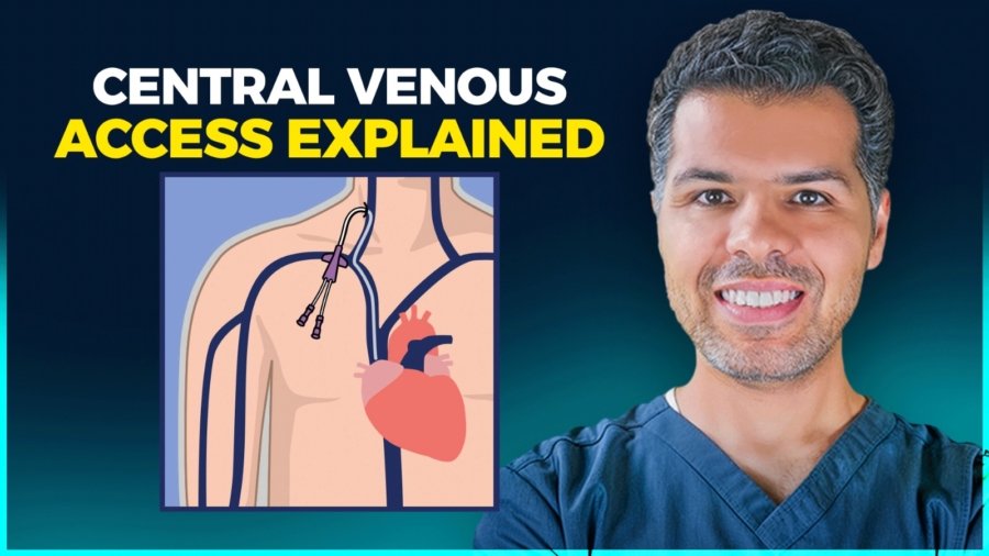
Includes short peripheral IVs (<6 cm) and midlines (8–20 cm).
Peripheral IVs: Inserted in hand or forearm veins for short-term use (3–5 days). Use feet only when upper extremity access isn’t possible.
Midlines: Placed in larger upper-arm veins for 1–4 weeks (up to 30 days max). They’re more durable but don’t reach central circulation.
Central access:
Catheters with the tip in central veins, classified as:
Non-tunneled Centrally inserted CVC:
These include internal jugular, subclavian, and femoral central lines.
They are used for short-term access — usually less than two weeks.
Subclavian is preferred for short-term use because it’s more comfortable and has a lower infection risk, but we avoid subclavian lines in CKD or ESRD patients because they can cause venous stenosis and compromise future dialysis access.
Femoral central lines should be reserved for emergencies or when the internal jugular or subclavian sites aren’t possible.
Two special types of non-tunneled central lines deserve mention:
Temporary dialysis catheters: These are large-bore central lines used when a patient needs urgent dialysis or CRRT. They’re usually placed in the right internal jugular or femoral vein if IJ access isn’t possible. If the need for dialysis becomes long-term, these should be transitioned to a tunneled dialysis catheter, like a Permacath.
Cordis — also called an “introducer sheath”: This is a short, large-bore central line that provides rapid access to the central circulation.
It’s used when we need fast fluid resuscitation, multiple infusions, or when placing a Swan-Ganz catheter in the ICU or OR. Cordis lines are meant for very short-term use — usually hours to a few days.
PICC line (Peripherally inserted central catheter): These catheters re placed in the arm and their tips end in the superior vena cava. Can stay for weeks–months (up to 6) for prolonged IV therapy, TPN, chemo.
Tunneled CVC (Hickman, Permacath): These are designed for long-term and frequent access — They run under the skin before entering the vein, which helps reduce infection risk. They’re commonly used for dialysis, parenteral nutrition, and ongoing chemotherapy.
Implanted Port (Port-a-Cath): These are fully under the skin. They’re accessed only when needed, require very little day-to-day care, and can last for years — often permanently — when maintained well.
2. Venous Accesses Ranking
By invasiveness from least to most:
Peripheral IV → Midline → PICC → Non-tunneled CVC → Tunneled CVC → Implanted Port
Prevent clogs: flush before / after meds + feeds, avoid mixing drugs, use warm water ± enzymes if blocked.
Introduction
An NGT, or nasogastric tube, is a flexible tube inserted through the nose, down the esophagus, and into the stomach, and sometimes down to the jejunum, also known as NJ tubes.
A gastrostomy tube is inserted directly through the abdominal wall into the stomach. The term PEG tube is often used to refer to any type of gastrostomy tube, though it technically describes those placed endoscopically (Percutaneous Endoscopic Gastrostomy). So from now on, PEG means all kinds of gastrostomy tubes.
Both deliver nutrition and medications, but the key difference is duration and invasiveness: NGTs are temporary, typically used for no longer than 4-6 weeks, while PEG tubes are meant for long-term feeding, usually more than four to six weeks, and allow patients greater comfort and mobility.
When should we consider NG tube placement in our patients?
Think about placing NGT tubes in the following situations:
Patients who can’t safely swallow and need medication and nutrition administration, such as mechanically ventilated patients, severe stroke patients, or any other reason.
Patients who need nutritional support when they cannot meet their caloric requirements through oral intake.
Patients who need gastric decompression for bowel obstruction or ileus.
NGT is a short term solution for up to 4-6 weeks, if the patient needs enteral support beyond this timeframe, it’s time to consider transitioning to a PEG tube.
PEG tubes are our go-to for long-term feeding—anything beyond four to six weeks. This includes:
Patients requiring prolonged mechanical ventilation who need to be transitioned to tracheostomy
Patients with prolonged dysphagia from neurological conditions like ALS, advanced dementia, or severe stroke.
It’s also indicated for head and neck cancer patients requiring radiation therapy, when we know they’ll need supplemental nutrition for more than four to six weeks.
And finally, when patients are heading home, and will need continued enteral feeding support.
Precautions and contraindications
NGTs are safe to place in most patients, and the absolute contraindications are limited. NGT placement is contraindicated in cases of facial trauma, nasal injury, abnormal nasal anatomy, or preexisting sinusitis. OGT (oral gastric tubes) can be alternatively considered in sedated patients, as it’s very uncomfortable to place in awake patients..
Patients with recent GI bleed (especially peptic ulcer with a visible vessel) are considered a relative contraindication. Gastroenterology should be consulted if an NGT is absolutely necessary here!
How about patients with liver cirrhosis? This is a frequently encountered situation! NG/OG tube placement is not absolutely contraindicated in cirrhotic patients! But, there is, of course, a higher risk of gastrointestinal bleeding, particularly in those with esophageal varices, particularly within 48 hours of NG tube placement. So, enteric tube placement should be considered only after failure of oral supplementation, and close monitoring for bleeding is warranted.
Percutaneous gastrostomy is strongly contraindicated:
in cirrhosis with ascites, due to high complication and mortality rates.
Active peritonitis, bowel ischemia, or mechanical intestinal obstruction.
Massive ascites and morbid obesity with a large panniculus are also recognized as major barriers to safe PEG placement.
Tube feeding system
NGT tubes come in different sizes, measured in French units. Common sizes range from 8 to 18 French. Smaller bore tubes, like 8-10 French, are more comfortable for patients and used primarily for feeding, while larger bore tubes, like 14-18 French, are used for gastric decompression. The tubes have depth markings along their length to help confirm proper positioning.
One of the most commonly used NGTs is the Salem sump tubes; these are large-bore tubes used mainly for gastric decompression and suction, featuring a double lumen with a blue vent to prevent mucosal damage.
The Dubhoff tube is a small-bore nasogastric feeding tube that is thinner and more flexible than standard nasogastric tubes, typically measuring 8-12 French in diameter. The tube features a weighted tungsten tip that helps it navigate through the gastrointestinal tract.
Choose small-bore soft tubes for nutrition and medication administration.
Choose large-bore, more rigid tubes for suction and gastric decompression.
In high-risk patients, such as liver cirrhosis or recent GI bleed, small-bore soft tubes are preferred and should be gently inserted.
PEG tubes consist of several parts: an external bolster or bumper that sits against the abdominal wall, a long tube shaft, an internal bumper that sits against the stomach wall, and a feeding port with a cap. Some PEG tubes have a balloon instead of an internal bumper. The key is that the tube creates a stable tract between the stomach and skin.
Gastrostomy tubes can be placed endoscopically (PEG), surgically, or by interventional radiology; as we said earlier, the term ‘PEG tube’ specifically refers to tubes placed by the endoscopic technique, but is commonly used for all.
PEG tube
Modern feeding systems include gravity bags, pump-controlled systems, and bolus syringes. Pump-controlled feeding is preferred for continuous feeds as it provides consistent delivery rates and reduces aspiration risk compared to gravity feeding.
NGT positioning confirmation
For NGT tubes, the gold standard is a chest X-ray showing the tube tip in the stomach, below the diaphragm, bisecting the carina. Bedside methods like pH testing and air insufflation are supplementary but should never replace X-ray confirmation for initial placement
Never use an NGT without X-ray confirmation
X-ray showing the tube tip in the stomach, below the diaphragm, bisecting the carina.
In this X-ray, the NGT tip is coiled in the esophagus and hasn’t entered the stomach; this needs repositioning
The X-ray shows that NGT has been inadvertently placed into the right mainstem bronchus.
For PEG tubes, we typically start using them within a few hours of insertion. In practice, we must get the okay from the physician who performed the procedure before using it. We must also inspect the site for proper positioning of the external bumper; it should sit flush against the skin but not too tight, as excessive tension causes pressure necrosis, while too much slack allows leakage.
A healthy PEG site with appropriate positioning.
This image shows buried bumper syndrome, where the internal bumper has migrated into the gastric wall, causing pain and feeding intolerance.
The daily bedside checks when rounding on patients with feeding tubes
Take all the necessary steps to minimize aspiration risk. Both NGT and PEG tubes carry aspiration risk, though PEG tubes generally have a lower risk of aspiration and aspiration pneumonia:
Maintain the head-of-bed (HOB) elevation at 30–45 degrees during and after feeding and avoid supine positioning whenever possible.
Pump-assisted continuous feeding is preferred over gravity or bolus feeds.
In high-risk patients such as those with previous aspiration or gastroparesis, post-pyloric or jejunal placement should be considered.
Inspect the insertion site for NGT/PEG:
Check the nares for pressure ulcers, and ensure the tube is secured properly at the correct depth marking.
For PEG: assess for redness, drainage, odor, or signs of infection. The site should be clean and dry. Also assess the pain or discomfort at the site.
Next, evaluate the patient’s tolerance: any nausea, vomiting, abdominal distension, or diarrhea? These suggest feeding intolerance.
Nurses routinely assess tube patency by gently flushing with water. Tubes should be flushed with 30 mL of water before and after each feeding or medication administration. If resistance is met, the tube may be clogged.
Routine residual checks in continuous feeds are no longer recommended except in select cases (e.g., high risk for intolerance or if it’s still part of the ICU protocols).
Tube maintenance and troubleshooting
For NGT
Clogged tubes are the most frequent issue. Prevention is key here: Flush with water regularly, crush medications, and never mix medications. If clogging occurs, try warm water flushes first. Pancreatic enzymes mixed with sodium bicarbonate can be used for stubborn clogs, but never use force, which can rupture the tube.
Tube migration or dislodgment needs immediate attention. For NGT, if the tube is pulled out partially, never advance it blindly; remove it completely and replace it with radiographic confirmation.
For PEG tubes
If the tube falls out within the first four weeks before the tract matures, this is an emergency requiring immediate replacement, ideally within hours, as the tract closes rapidly. After four weeks, the tube can be replaced at the bedside with a Foley catheter temporarily until a new PEG tube is placed.
Residual volume troubleshooting (If you decided to check residual volume in continuous feed)
Remember that metoclopramide and erythromycin can be given every 6 hours as needed and remember that both can prolong QTc.
If intolerance persists despite prokinetics, switch to post-pyloric feeding
PEG site infections
Mild cellulitis around the site responds to oral antibiotics and local care. Severe infections, abscess formation, or necrotizing fasciitis require surgical consultation.
Buried bumper syndrome
presents with feeding intolerance, pain, and difficulty flushing. This requires endoscopic or surgical intervention to reposition or replace the tube.
A chest tube is a hollow, flexible tube that we place into the pleural space to drain air, blood, or fluid so the lung can re-expand.
A pigtail catheter is simply a smaller, softer version of a chest tube, with a curled ‘pigtail’ tip.
Both do the same job — removing air or fluid from around the lung — but the big difference is in size and invasiveness.
Indications
This table summarizes the indications of the traditional chest tube vs the pigtail catheter
The chest tube drainage unit
An adequate chest drainage system aims to:
Remove pleural fluid and/or air.
prevent their reflux into the pleural space.
Restore negative pleural pressure (less than atmospheric pressure) to allow lung re-expansion.
To understand how the chest tube achieves that, we must understand the components of the chest tube drainage unit.
The one-bottle unit
Back in the day, the chest tube unit was simply a tube connected to a bottle filled with saline/water and a one-way valve. While this works well for draining air, it doesn’t for fluid! If a pleural effusion is drained, the fluid level in the bottle will quickly increase, reducing the efficiency of removing additional fluid from the patient
The two-bottle unit
To solve this problem, a second water seal bottle was introduced! This leaves the first bottle for collection only and one for the water seal.
To understand the water seal, try blowing some air in a cup of water. You see all these bubbles; can you suck that air back? Of course, you can’t! The air gets sealed in the water.
The three-bottle system
To improve drainage efficiency, a third bottle connected to external suction is added. a collection bottle, a water seal bottle, and a suction bottle.
The modern chest tube unit
Nowadays, all these bottles/chambers are currently integrated into a single modern, multifunctional, easy-to-manage unit.
The collection chamber, the water seal chamber, and the suction chamber. Notice the air leak monitor!
The tube itself comes in different sizes and shapes. They can be straight, angled, spiral, or coiled at the end (referred to as “pig-tail”).
Confirmation of proper positioning with CXR
Ideally, the tip of the chest tube should be positioned at the apex if inserted for pneumothorax, and at the base if inserted for fluid removal. Ensure the tip is not outside the pleural space or abutting the mediastinum.
A chest tube was inserted for pneumothorax and is in good position toward the apex.
A pigtail catheter at the lung base draining fluids
A pigtail is abutting against the mediastinum and should be pulled back
One of the chest tube fenestrations is outside the pleural space! It should be inside.
The fenestrations seen on chest tubes are these evenly spaced gaps (interruption marks) along the radiopaque stripe of the tube. The fenestrations should be inside the pleural space to allow effective drainage of air or fluid while reducing the risk of clogging or blockage. Multiple holes also distribute suction evenly, preventing tissue trauma and ensuring continuous tube function.
The daily bedside checks on patients with chest tubes
Review the CXR first.
Although a daily CXR isn’t necessary in patients with chest tubes who are stable, it must be obtained if there is a change in the patient’s clinical status, after major adjustments, repositioning, or removal of the chest tube, or if there is a persistent air leak or fluid drainage.
Assess the output in the collection compartment. Is it bloody or not? Is it increasing or decreasing?
Check for tidaling! Tidaling is the rise and fall of fluid in the water seal chamber with breathing. Watch this YouTube video showing tidaling!
Tidaling tells you the chest tube is working and still in contact with the pleural space. If tidaling disappears, it means one of two things:
The lung has fully re-expanded — which is good —
The tube is blocked — which is bad —
If the patient’s breathing comfortably, chest X-ray shows full lung expansion, and there are no signs of distress — that’s good. But if the patient is short of breath, there’s new subcutaneous emphysema, or the tube is kinked or clogged — that’s bad, it means obstruction.
Check for bubbling! Do you see any air bubbles in the air leak monitor? Check bubbles in the air leak monitor in this YouTube video:
Bubbling means air is still escaping. It could be from an ongoing air leak in the pleural space or a disconnection in the tubing outside the chest.
Never clamp a chest tube if bubbling is present, please! If you do, tension pneumothorax can develop quickly.
Check the dressing and assess the tube integrity from the dressing to the tube unit, particularly if you see a continuous bubbling in the air leak monitor.
Assess the Pain level! Chest tubes, especially large-bore ones, are painful, so please ensure adequate pain control.
Ask them to ambulate. Chest tubes don’t mean they can’t be ambulated.
Do they still need the chest tube?
Chest tube removal
The chest tube should only be removed by the team that placed and is actively managing it. Typically, the ICU, thoracic, cardiovascular, or trauma surgery team.
Removal is considered when:
The active issue has been resolved, whether fluid or air.
The lung has fully expanded.
The patient is clinically stable.
For example, in cases of draining fluid, the amount in the collection compartment is consistently low; something like less than 20 cc/day for 1-2 days.
And for pneumothorax, you don’t see any air bubbles in the air leak monitor for 1-2 days, and the lung has fully expanded.
Typically chest tube is clamped for 12-24 hours (clamping deems the tube not functioning). If the patient’s conditions remained clinically stable with a stable CXR, then this means it’s time to remove the tube.
DO NOT CLAMP CHEST TUBE on your own unless you are part of the team who is actively managing it. DO NOT EVER CLAMP A TUBE IF YOU SEE AIR BUBBLES IN THE AIR LEAK MONITOR!
Some patients may need the chest tube for weeks and months, like malignant effusions. In such a case, a traditional chest tube can be replaced with a pigtail catheter to go home with.
Pitfalls & Warnings
Don’t connect the chest tube to suction immediately after insertion to reduce the risk of re-expansion pulmonary edema. Re-expansion pulmonary edema is a non-cardiogenic edema that happens when a lung that’s been collapsed for days is rapidly reinflated — the fragile capillaries leak, flooding the alveoli. Management is supportive with oxygen, noninvasive ventilation, and cautious fluids. The real key is prevention: never drain too much, too fast.
Diuretics may worsen hypovolemia and are not recommended. The re-expansion pulmonary a non-cardiogenic edema driven by increased capillary permeability from reperfusion injury, not by fluid overload
Watch for a blocked/clogged tube (no drainage but effusion enlarging). Sometimes, a thrombolytic is used for clogged tubes!
Learn the 8 bad lab habits in hospitals that waste resources and rarely change management. Discover smarter, evidence-based alternatives to improve patient care and reduce unnecessary testing.
I can’t remember the last time I finished a shift without a call about a patient in acute pain. In this post, I’ll share a practical, easy-to-use guide to help you choose the right analgesia for your patients.
Pain severity scale
The first and most important step—often overlooked—is to assess the severity of pain using a standardized pain scale. In the hospital, this is typically a numerical rating scale, where patients are asked to rate their pain from 0 to 10. A score greater than 6 is generally considered severe.
Mild pain
This is pretty straightforward. Oral acetaminophen alone or in combination with a low-dose oral NSAID can be used. An ideal mild pain regimen would consist of:
Acetaminophen 650 mg orally every 4 hours as needed.
Ibuprofen 400 mg orally every 4 hours as needed.
The nurse should administer either option, but not both at the same time. They may alternate between the two if needed.
Personally, I prefer the sole use of acetaminophen in the mild pain category.
Moderate pain
A high-dose Oral NSAID with or without low-dose IV acetaminophen.
IV NSAIDs are an option here, as well. In such a case, oral NSAIDs should be discontinued. An ideal regimen would consist of:
An NSAID (Pick one):
Naproxen 500 mg orally twice daily as needed.
Ibuprofen 600 mg orally every 6 hours, or Ibuprofen 800 mg orally every 8 hours as needed.
Ketorolac 15 mg IV every 6 hours as needed.
If NSAIDs are contraindicated or inadequate, Acetaminophen 650 mg IV every 6 hours as needed can be added.
“Don’t use oral and IV NSAIDs together”
Severe pain
This is the category where opioids should be used! Opioids should be reserved for severe pain only, including oral & IV opioids! This may seem odd, as a lot of hospital pain protocols include low-dose oral and IV opioids for moderate pain. Please avoid that unless there is a contraindication to use acetaminophen and NSAIDs!
Why am I saying this?
Adding opioids for moderate pain will likely lead to unnecessary and avoidable use of opioids!
Just imagine your patient is having moderate pain based on the assessment scale, and you have the option to pick between a low-dose opioid or an NSAID, most of us will unconsciously lean toward picking the stronger analgesic, which is an opioid here, although a dose of naproxen would have been probably adequate!
So, unless there is a contraindication to use NSAIDs or Acetaminophen, reserve opioids for severe pain only, please.
But does that mean opioids are the only option for severe pain? Of course not!
High-dose IV Acetaminophen and IV NSAIDs are pretty effective in severe pain as well.
So an ideal regimen for severe pain analgesics would consist of:
IV Acetaminophen or IV NSAIDs (First line)
Oral opioid (Second line)
IV opioid (Last to use).
I will go for IV NSAIDs or Acetaminophen first, oral opioids second, and IV opioids are our last resort if the patient’s acute pain remains inadequately controlled
Remember that NSAIDs are first-line analgesics in the following conditions:
Renal colic, IV Ketorolac in particular, is effective here.
Musculoskeletal pain like arthritis pain, pleuritic-type chest pain, rib fracture, and muscular strains.
Incisional post-operative pain.
Acetaminophen is particularly effective in most types of headaches alone or in combination with caffeine.
Also, the location of pain may warrant analgesia avoidance, like chest pain of cardiac origin, where NTG should be used instead.
Now, which opioid should we use and what dose? I refer you to an older post where I discussed the use of opioids. You can read it here
Persistent severe pain
In persistent severe pain where the patient keeps requiring recurrent doses of opioids despite treating or while treating the underlying cause of pain, we need to try to reduce the pain level with other means and consequently reduce the need for IV opioids:
Adding scheduled (Not PRN) oral acetaminophen or oral NSAID, remember to discontinue their IV equivalents.
Adding Gabapentin
Adding a scheduled low-dose oral opioid can be considered if previous measures didn’t help reduce the use of IV opioids.
Let me give you an example:
A 64-year-old lady was admitted with a pelvic fracture, she’s in severe pain her severe pain regimen consists of the following:
Ketocorolac 15 mg IV every 6 hours as needed
Oxycodone/acetaminophen 5/325 mg PO every 4 hours as needed
Morphine 4 mg IV every 6 hours as needed
You reviewed her chart and found that she received 12 doses of IV morphine over the last three days, her nurse reported that IV morphine is the only thing keeping her pain under control and she asked you if there’s anything we could do to help her pain control.
Of course, the first thing I do is check if the patient has received IV Ketorolac and oxycodone/acetaminophen or if the nurse just jumped to IV morphine, if they have not been tried, I will ask the nurse to try them first and assess the response! If they have already been given with inadequate response, then we can do the following:
Add scheduled high-dose oral ibuprofen, or naproxen, or acetaminophen
Add Gabapentin
If that remains inadequate, we add a scheduled oral opioid for a few days while escalating the Gabapentin dose
Please remember this about the use of Acetaminophen and NSAIDs:
Don’t combine oral and IV forms of the same medication or the same class within the same severity category, for example, don’t use oral naproxen and IV ketorolac together for moderate pain, or oral acetaminophen and IV acetaminophen together for severe pain.
Combining acetaminophen at doses higher than 2 g/day together with an NSAID isn’t more effective than an NSAID alone and might be associated with a higher risk of gastrointestinal complications! So keep that in mind!
The total daily amount of acetaminophen shouldn’t exceed 4 g/day including all forms of acetaminophen, whether oral or IV, and the one combined with oral opioids.
More options for severe pain
There are other options to consider in severe pain cases:
Regional block performed by surgery or anesthesia can be helpful in rib fractures or postoperative pain
Ketamine can be considered in severe pain refractory to opioids.
Dexamethasone can be considered where swelling and edema are suspected as a contributing factor to the pain line in disk herniation or metastatic bone pain.
Local anesthetics like lidocaine patches are not that effective in acute severe pain management, but they can be used as an adjuvant therapy to other analgesics for superficial musculoskeletal pain.
Muscle relaxants can be considered when muscle spasms may contribute to the patient’s acute pain
In this post, I will share when to use them, how to use them, what to monitor, and their alternatives:
(1) IV Vancomycin
In the ICU, I reach for IV vancomycin when MRSA or resistant gram-positives are a concern — like sepsis of unknown source, ventilator pneumonia, line infections, or resistant Strep pneumo meningitis. It’s my go-to until cultures guide me otherwise, with close drug monitoring and quick de-escalation when I can.
The initial loading dose is 20-35 mg/kg, based on actual body weight, and should not exceed 3,000 mg. So, for a 70 kg patient, it’s roughly 1.5-2 gm. Do not renally adjust for the loading dose!
Subsequent dosing is guided by the area under the concentration-time curve to minimum inhibitory concentration ratio (AUC/MIC), not by trough-only monitoring. Fortunately, our pharmacists take care of that.
Vancomycin can cause nephrotoxicity and agranulocytosis, which is why we monitor kidney function and CBC while on IV vancomycin.
Vancomycin infusion reaction (previously “Red man syndrome”) occurs with IV administration. It’s a side effect, not a true allergy. Manage by slowing the infusion rate to 50% or 10 mg/min (whichever is slower). Diphenhydramine plus famotidine effectively relieves symptoms. Stop infusion only for hypotension, chest pain, dyspnea, or muscle spasms; restart at reduced rate when resolved.
Remember that Oral vancomycin is solely used for C. diff treatment.
If I can’t use IV vancomycin because of allergy or toxicity, Linezolid and daptomycin are the main alternatives, and the source of infection dictates which is better to pick:
Linezolid → preferred for lung, CNS, and skin/soft tissue infections. Easy, weight-independent dosing, 100% oral bioavailability, and safe in renal failure. But it’s bacteriostatic, not ideal for bacteremia, and can’t be combined with serotonergic drugs due to serotonin syndrome risk. Limit use to ≤28 days because of thrombocytopenia, neuropathy, and myelosuppression.
Daptomycin → preferred for bloodstream infections, endocarditis, and other deep-seated infections (MRSA or VRE). Bactericidal, but weight-based and requires renal adjustment. Never use it in pneumonia — it’s inactivated by surfactant. Rhabdomyolysis and eosinophilic pneumonia are potential serious side effects of daptomycin.
Ceftaroline → the only β-lactam with MRSA activity. I reach for it when linezolid or daptomycin can’t be used or when the vancomycin MIC is elevated (≥2 μg/mL). It’s useful for pneumonia and bacteremia, but requires renal dose adjustment.
Linezolid vs Daptomycin vs Ceftaroline
(2) Cefepime
I reach for cefepime when broad-spectrum coverage of Gram-negative and Gram-positive bacteria is required, such as in suspected hospital-acquired or ventilator-associated pneumonia, and sepsis of unknown source.
That’s why you see quite often the combination of vancomycin and Cefepime in initial empiric treatment for severe infection and sepsis. Vancomycin to cover for MRSA and cefepime for the rest, including Pseudomonas. Cefepime lacks anaerobic coverage, and metronidazole should be added if anaerobic coverage is required.
Cefepime has good CNS penetration and can be used to treat CNS infections.
Cefepime isn’t effective against extended-spectrum beta-lactamase (ESBL) producing organisms, regardless of culture and susceptibility results. Use carbapenems instead for that.
Dosing is 1-2 gm IV every 8-12 hours and should be adjusted to renal function.
Monitor mental status to detect cefepime neurotoxicity early. Neurotoxicity in the form of encephalopathy, confusion, hallucinations, stupor, coma, aphasia, myoclonus, seizures, and nonconvulsive status epilepticus can all happen particularly with the use of high doses in patients with renal impairment.
If penicillin allergy is reported in the patient’s chart, ask what kind of allergy. Cefepime should only be avoided in patients who report a history of immediate hypersensitivity reactions to penicillins (such as anaphylaxis, angioedema, or urticaria).
(3) Piperacillin/Tazobactam (Zosyn)
In the ICU, I reach for Piperacillin/Tazobactam when broad-spectrum coverage of Gram-negativeincluding Pseudomonas, Gram-positive, and anaerobic bacteria is required (No need to add metronidazole).
It was widely used in combination with vancomycin for the initial empiric antibiotic coverage for severe infections until it was found that this combination increases the risk of AKI! Since then, it has been largely replaced by cefepime.
It has poor CNS penetration and shouldn’t be used for CNS infections.
Dosing of (piperacillin and tazobactam) is dependent on the type and severity of infection being treated, and it requires renal dosing adjustment.
(4) Meropenem
In the ICU, I reach for meropenem when ESBL-producing organisms are suspected, or when cefepime and piperacillin/tazobactam can’t be used due to a serious allergy.
It covers Gram negatives, Gram positives, and anaerobes, and it has excellent CNS penetration, making it suitable for CNS infections.
It has a very low cross reactivity with penicillin’s making it a great alternative in patients with serious penicillin allergy, even in those with a history of immediate hypersensitivity reactions (such as anaphylaxis, angioedema, or urticaria).
Among carbapenems, ertapenem lacks pseudomonal coverage.
Cefepime vs Piperacillin/tazobactam vs Meropenem
(5) Bactrim (Trimethoprim–sulfamethoxazole)
The reason I include it here because it’s the antibiotic of choice to resort to when thinking about PJP pneumonia, Nocardia, and in the treatment of stenotrophomonas maltophilia, an opportunistic gram-negative bacillus that is a common culprit in hospital-acquired infections in critically ill patients.
Dosing is weight-based. The IV preparation of Bactrim entails the use of a large volume of dextrose solution to mix with, so watch for the risk of hyponatremia.
Renal adjustment is required; we need to closely monitor CBC, kidney function, and electrolytes as hyperkalemia, bone marrow suppression, and hyponatremia are all potential side effects of Bactrim.
Allergy to nonantibiotic sulfas isn’t a contraindication to use sulfa antibiotics.
Conclusion
I know there are many other important antibiotics I didn’t cover today. I wanted to keep this post focused and concise. If you’d like a bit deeper practical dive into the antibiotics we use most often in clinical practice, check out my eBook: Antibiotics in Real-Life Clinical Practice: A Pocket Guide for Residents, Hospitalists, Internists, Nurse Practitioners, and Physician Assistants. Check it out here
Mechanical ventilation doesn’t have to be intimidating. If you are a non-ICU doc who just wants to feel more prepared when your patient gets intubated, this post is for you! I’m pretty sure after this, you won’t shy away from the ventilator again.
So grab a coffee, and let’s finally make sense of mechanical ventilation. Let’s get started.
Concept (1): The respiratory cycle has two phases: expiration and inspiration.
Inspiration is an active process that requires energy. Expiration is typically passive, relying on the lungs’ natural elastic recoil.
The respiratory cycle duration is calculated by dividing 60 seconds by the respiratory rate (RR). This cycle encompasses both inspiratory and expiratory time. The relationship between inspiration and expiration is not equal; physiologically, the ratio of inspiratory time to expiratory time (I:E) is typically 1:2 or 1:3, with expiration being longer than inspiration.
Respiratory cycle and I:E ratio
Example:
RR = 20 breaths/minute
The respiratory cycle duration = 60/20 = 3 seconds
Using I:E ratio of 1:3, the inspiratory time would be 0.75 seconds, and the expiratory time would be 2.25 seconds.
Concept (2): The ventilator does the heavy lifting during inspiration, but it doesn’t clock out during expiration!
As we mentioned earlier, inspiration is an active process that requires energy, so the ventilator works mainly during this active, energy-consuming phase. But that doesn’t mean the ventilator just shuts off during expiration—it continues to play a crucial role by maintaining a certain level of pressure that is called PEEP, or Positive End-Expiratory Pressure.
PEEP prevents the alveoli from collapsing at the end of expiration by holding a small amount of pressure in the lungs throughout the entire respiratory cycle, not just during the expiration phase. Think of it like a doorstop that keeps the alveoli slightly open, making the next breath easier and improving oxygenation.
Pressure vs time graph on a ventilator screen. The yellow line represents the PEEP, and the green line represents the inspiratory pressure that is added on top of the PEEP.
Don’t be fooled by the name—PEEP isn’t just at the end of expiration. It’s present throughout the entire respiratory cycle. During expiration, it’s the only pressure applied, and during inspiration, the ventilator adds pressure on top of the PEEP to deliver the breath.
Concept (3): Air flows from higher-pressure to lower-pressure areas.
During inhalation, air enters the lungs because the pressure at the nose and mouth is higher than in the alveoli. During exhalation, the pressure in the alveoli becomes higher than at the airway opening, so air flows out. This air movement continues as long as a pressure gradient exists. In spontaneous breathing, we actively lower the pressure inside the alveoli below atmospheric pressure, creating suction and pulling air in.
Air flows from higher to lower pressure areas
In mechanical ventilation, the ventilator does the opposite: it raises the pressure at the nose and mouth above alveolar pressure to push air in. That’s why we call it a positive pressure breath
In spontaneous breath, we suck the air in; while in positive pressure breath, the ventilator pushes the air in.
Concept (4): To get air in, the ventilator has to fight three battles: the tube, the airways, and the lungs.
During mechanical ventilation, the ventilator delivers air through the endotracheal tube (ETT), down the airways, and into the alveoli. To do this, it must overcome two main types of resistance:
The airway resistance (from the ETT and conducting airways).
The elastic resistance (from the alveoli and lung tissue).
The airway resistance
This resistance is primarily due to their narrow lumens and flow dynamics. The pressure required to overcome this kind of resistance is called Resistive pressure, and it is calculated by multiplying the flow by the airway resistance.
Resistive Pressure = Flow (F) × Resistance (R)
The elastic resistance
The alveoli are evaluated based on their compliance—their ability to stretch and accommodate volume. The pressure required to overcome alveolar and lung tissue stiffness is called Elastic pressure, and it’s calculated by dividing volume by compliance.
In mechanical ventilation, the volume here simply represents TV, and the elastic pressure represents the alveolar pressure, so the equation can be rewritten as:
Alveolar pressure = TV/C
Therefore, for a full breath to be delivered, the ventilator must generate enough total pressure to overcome both the resistive and elastic or alveolar pressures. In mechanical ventilation, this pressure is called the peak inspiratory pressure (PIP).
PIP is proportionally related to Flow, airway resistance, and TV.
Inversely related to compliance.
The relation between PIP, the flow, airway resistance, TV, and compliance
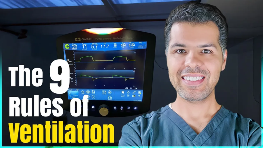
Take a peek at this ventilator screen: The PIP is 20 cmH2O, the flow rate is 44 L/min, and the tidal volume is 500 mL.
PIP is 20 cm H2O, which means 2o cmH2O was required to overcome the airway and elastic resistance and deliver this breath.
Guess what will happen to the PIP if I increase the flow or the tidal volume? Based on the equation, the peak inspiratory pressure (PIP) will increase.
What will happen to the PIP if the patient bites the endotracheal tube or develops bronchospasm, both of which increase airway resistance? Again, you’ll also see a rise in PIP.
Let’s say the patient develops pulmonary edema and bilateral pleural effusion. What would happen to the PIP in such a case? Pulmonary edema and bilateral pleural effusion reduce the alveoli’s ability to accommodate tidal volume (reduce their stretchability and compliance). PIP is inversely related to compliance; now the ventilator has to push harder to get the breath in, which means higher PIP is required!
Concept (5): Plateau Pressure: Your Window into Lung Compliance
Let’s imagine a moment when the flow drops to zero— this occurs briefly at the end of inspiration, just before expiration begins, and again at the end of expiration, just before the next inspiration starts. These are the transition points between phases of the respiratory cycle when there’s no airflow because the pressures between the airway and alveoli are equal.
Guess what happens to the PIP when the flow drops to zero? Let’s go back to our equation:
If the flow drops to zero, the resistive component disappears, and the equation simplifies to:
PIP = TV/Compliance = Alveolar Pressure.
This means when the flow is zero, the PIP is equal to the alveolar pressure, and this makes sense as pressure gradient means flow, and the absence of the gradient means no flow.
In mechanical ventilation, the pressure measured at the end of inspiration when the flow ceases is known as the Plateau pressure. Plateau pressure provides great insight into lung compliance. The lower the plateau pressure, the more compliant, stretchable, and less stiff the lung is, and vice versa!
Plateau pressure is our eye into lung compliance
How do we measure the plateau pressure? Plateau pressure is measured using the inspiratory pause or hold maneuver. In clinical practice, we aim for a PIP < 35 cmH2O and a plateau pressure of < 30 cmH2O.
How about the opposite? I mean the pressure measured at the end of expiration and before the inspiration valve opens? What do you think the alveolar pressure should be in such a case? We mentioned earlier that PEEP is the only pressure the ventilator provides during expiration, which means the expiratory flow will continue until the alveolar pressure drops from the maximum value at the end of inspiration (plateau pressure) down to the PEEP value, when the flow ceases. So normally, the alveolar pressure measured at the end of expiration when there is no flow must equal PEEP.
To confirm that, we perform what we call the expiratory pause maneuver. This maneuver allows us to check for any gas trapping and autoPEEP.
Concept (6): The ventilator can’t think for itself—it needs our guidance
For the ventilator to function properly, we have to give it five instructions:
When to start the breath.
How to deliver the breath.
When to terminate the breath.
How much oxygen to give?
How much PEEP to give?
1. When to start the breath or inspiration?
The ventilator listens to the patient to detect the patient’s inspiratory effort. It will pick up any inspiratory effort as a sign that the patient is trying to breathe, and it will assist the patient with that breath! This is known as a patient’s triggered breath or, as we call it in mechanical ventilation, an Assist breath or “A” breath.
But what happens if the ventilator doesn’t detect any patient trigger? Let’s say the patient is paralyzed or brain-dead.
In this case, the ventilator has a built-in safety mechanism: it will deliver a breath if no patient’s trigger is detected for a specific time! This is called a backup breath or, as we call it in mechanical ventilation, a Control breath or “C” breath.
Take a look at these two ventilator screens. The breath on picture A is a controlled or backup breath labeled as C breath, and the breath on picture B is an assist breath triggered by the patient and assisted by the ventilator.
Demonstration of (A) & (C) breaths
But what decides how long the ventilator should wait before delivering a backup breath? This timing is based on the set respiratory rate (RR), which is known as the backup rate. The backup rate is the minimum number of breaths/minute the patient receives, even if they make no effort to breathe. The ventilator will listen to the patient for a period equal to [60 sec/backup RR] before delivering the backup breath.
Example: If the backup rate is set to 15 breaths per minute, the ventilator waits up to 4 seconds (60 ÷ 15), and if no patient’s trigger is detected, a backup breath will be delivered!
The actual RR is either equal to or higher than the backup rate, but never lower! This is important when trying to adjust the backup RR. Let’s say the backup rate is 14 and the patient is breathing at 20 breaths/min; there is no point here if you increase the backup rate to 16 because the actual RR is already higher!
Take a look at this ventilator screen, see the set backup rate at 18 while the actual RR on the top is 28. Increasing the backup rate will not have any impact unless it becomes higher than the actual RR.
The actual RR at the top is 28, while the backup RR is at the bottom set at 18. The pieces of information at the top are a live transmission of the actual reading. Values at the bottom are the set values.
The values at the bottom of the picture are the set values that we provide, while those at the top are the actual live values! The ventilator tries to fulfill the set values or close to them (See the set TV is 500, but the actual one is 524).
Back to the trigger, how does the ventilator detect the patient’s trigger?
In spontaneous breathing, the patient generates negative pressure in the alveoli, which creates a suction effect that pulls air in. When the patient is intubated and connected to a ventilator, this same effort draws air through the circuit, and that’s how the ventilator knows it’s time to assist.
Take a look at the ventilator circuit picture below: you’ll notice two limbs connected to the endotracheal tube (ETT)—the inspiratory limb, which carries air from the ventilator to the patient (often passing through a humidifier), and the expiratory limb, which returns air from the patient back to the machine.
When the patient is not making any effort, air flows equally through both limbs. When the patient initiates a breath, he pulls some air from the inspiratory limb into his lungs, reducing the flow returning through the expiratory limb. The ventilator senses this imbalance and interprets it as an inspiratory effort and will go ahead and assist the patient with that breath. This is called a flow trigger.
Alternatively, the ventilator can detect a drop in airway pressure (caused by the patient’s inspiratory effort) and use that as a trigger. This is called a pressure trigger.
🔁 To summarize: the ventilator can recognize a patient’s attempt to breathe in two ways:
Flow Triggering – by sensing a drop in flow through the expiratory limb.
Pressure Triggering – by sensing a drop in airway pressure.
Take a look at these two screenshots of two ventilators:
Pressure vs flow trigger
On Picture A, Psens shows the pressure trigger. It’s set at 2 cm H₂O, meaning the patient must reduce his airway pressure by at least 2 cm H₂O to trigger a breath. We usually set this between 2 and 3 cm H₂O.
On picture B, Vsens represents the flow trigger, set at 3 L/min. This means the patient must draw in at least 3 L/min (approximately 50 mL/sec) for the ventilator to deliver a breath.
Whether using a flow trigger or a pressure trigger makes no clinical difference—neither offers a clear advantage over the other.
2. How should the ventilator deliver this breath?
In mechanical ventilation, we refer to the way of delivering the breath as the “target.” Although the term can be confusing, the concept is straightforward—just give me your full attention!
Let’s bring back our lovely equation again:
PIP = (F x R) + (TV/C).
Which can be rewritten as: PIP = (F x R) + Alveolar pressure. Which can be rearranged into: F = (PIP – Alveolar pressure) / Resistance.
In mechanical ventilation, we can directly set the flow rate and the peak inspiratory pressure (PIP) on the ventilator. However, alveolar pressure and airway resistance are not directly settable—they’re influenced by the patient’s lung mechanics and underlying condition.
Based on the equation, mathematically, for the pressure–flow relationship to hold true, we can only set one variable—either the pressure or the flow, but not both. That’s because one must remain flexible to allow the other to stay fixed. If we fix both, the equation wouldn’t hold under changing patient conditions.
This will leave us with one of two options to deliver the breath:
You can either set the flow and let the pressure adjust, which is called the flow target.
Set the pressure and let the flow adjust, which is called the pressure target.
In flow-targeted ventilation, we start by setting the desired flow rate. From there, we have two main subtypes based on how that flow is delivered:
We can ask the ventilator to keep the flow rate fixed throughout the inspiratory phase, which results in a square or rectangular flow vs time curve.
Or we can ask the ventilator to give the set flow rate only at the beginning of the breath, and let it progressively drop or decelerate throughout the rest of the inspiratory phase! This results in a decelerating ramp flow vs time curve.
In clinical practice, we primarily use the decelerating ramp flow, it generally results in a lower peak inspiratory pressure (PIP) compared to the constant (square) flow pattern.
Take a look at this flow target on this ventilator screen and notice:
The green curve shows the decelerating ramp flow pattern.
On the info panel, the flow target is set at Vmax: 44 L/min with the ramp type set to decelerating.
Decelerating flow pattern and Vmax.
How about the pressure target ventilation?
Here, we tell the ventilator to apply a fixed amount of pressure on top of the PEEP throughout the inspiratory phase until the breath is delivered.
What do you think the flow pattern will be in the pressure target? To maintain a fixed pressure throughout the inspiratory phase, the flow must progressively decrease to keep this equation valid, “F = (PIP – Alveolar pressure) / Resistance”. Of course, as long as the resistance is constant. That’s why the flow vs time curve is always a decelerating ramp curve in pressure target modes.
Take a look at this ventilator screen, see the fixed pressure here. The ventilator adds 20 cmH2O on top of PEEP of 10 and keeps it there during the whole inspiration. The flow curve is a decelerating ramp curve.
Pressure target ventilation
What do you think will happen to the flow rate if we set the inspiratory pressure (Pi) at 30 cmH2O instead of 20?
If we increase the inspiratory pressure from 20 to 30 cmH₂O, the flow rate will increase; so the higher the set pressure, the greater the initial flow the ventilator delivers.
3. When should the ventilator stop delivering the breath (Cycle)?
This is known as the ‘cycle’ in mechanical ventilation. There are three main mechanisms to determine when the ventilator stops inspiration and switches to expiration. These are known as cycling mechanisms:
Volume-Cycled Ventilation:
In volume-cycled breaths, the ventilator ends inspiration once the preset tidal volume (TV) is delivered.
🔍 Look at the ventilator screen below and see the volume vs time curve. Once it reaches the desired TV, it will switch from green color, which represents inspiration, to yellow color, which represents expiration. Also, look at the top panel and see that the actual TV was 315! In reality, the ventilator doesn’t always fulfill the exact set TV, but it will be close to that!
Volum-cycled ventilation
What do you think will happen to the inspiratory time if we increase the TV here? Of course, it will increase; the larger the volume, the longer it takes to deliver it. The opposite is true.
Time-Cycled Ventilation:
In time-cycled breaths, the ventilator switches to expiration after a preset inspiratory time has elapsed, regardless of the volume delivered.
🔍 Check the ventilator screen for the set inspiratory time and see that the green line in all the curves switches to yellow after the set inspiratory time.
What do you think the TV will be if we shorten the insp time? It will be smaller as we spending less time delivering the breath, the opposit is true.
Flow-Cycled Ventilation:
In flow-cycled breaths—used primarily in pressure support mode—the ventilator ends inspiration when inspiratory flow drops to a preset percentage of peak inspiratory flow.
🔍 Look at this screen and see the “Esens” or expiratory sensitivity setting (commonly set at 25%)—the breath ends when flow falls to 25% of its peak during inspiration.
What will happen to the TV if we set ESens at 50% instead of 25%? It will be smaller as we are having a shorter inspiratory time! The opposite is true.
4. How much FiO₂ and how much PEEP? These two are oxygenation parameters, and we’ll discuss them later in this post.
Concept (7): Every Mode Is a Recipe: You Just Change the Ingredients
At the heart of mechanical ventilation are three fundamental modes:
🔹 Volume Control.
🔹 Pressure Control.
🔹 Pressure Support.
Every other mode you’ll encounter is either a hybrid or a modification of these three.
To make sense of any mode, just break it down into the three key ingredients we just explained:
🟢 Trigger – What starts the breath?
🟡 Target – How to deliver the breath?
🔴 Cycle – What ends the breath?
Before I go further!
If you’re finding this helpful, you’re going to love my full mechanical ventilation course. It covers everything we’re walking through in this video—from triggers, targets, and cycling to all the core modes—plus much more, like: Troubleshooting PIP, Rise time, Mean airway pressure, Auto-PEEP and gas trapping, Assessing weaning readiness, and how to do a proper weaning trial.
It’s designed to walk you through mechanical ventilation step-by-step, in a way that’s clear, visual, and practical.
If you’re ready to truly master the vent and feel confident at the bedside, check out the course.
Volume control or VC mode
Trigger: VC mode typically uses assist/control (A/C) triggering. As we explained earlier, the ventilator delivers a breath either when the patient initiates one or, if the patient doesn’t, the ventilator will trigger a breath based on the set backup respiratory rate.
Target: VC mode is flow-targeted, commonly using a decelerating ramp flow pattern.
Cycle: VC is volume-cycled, meaning the breath ends once the preset tidal volume (TV) has been delivered.
So when ordering VC mode, in addition to the FiO2 and PEEP values, we need to provide the respiratory therapist with the following values:
The backup RR.
The TV.
The Flow curve: square or decelerating.
The Peak flow rate.
In most cases, RT will, by default, pick a decelerating ramp flow curve and set the peak flow rate; a reasonable starting peak flow rate is between 40-60 L/min.
Take a look at this VC mode screenshot,
VC mode with A/C trigger, and that specific breath was a control breath. The backup rate is set at 18, this is the minimum RR the patient will have. The target is a flow target with a peak flow rate set at 44 L/M with a decelerating ramp, the breath will terminate once a TV of 500 cc is delivered.
Pressure Control (PC) Mode
Trigger: Like VC mode, PC mode uses assist/control (A/C) triggering, meaning the breath can be triggered either by the patient’s effort or by the ventilator’s set backup respiratory rate if no spontaneous inspiratory effort is detected.
Target: As the name suggests, PC mode is pressure-targeted. The ventilator delivers a breath by applying a set inspiratory pressure above the PEEP, and adjusts flow as needed to maintain that pressure throughout inspiration.
Cycle: PC mode is time-cycled—the breath ends when the set inspiratory time has elapsed, regardless of how much volume was delivered.
When ordering PC mode, in addition to the FiO2 and PEEP values, we need to provide the respiratory therapist with the following values:
Back up RR.
Pi or inspiratory pressure, this is the pressure that will be added on top of the PEEP during inspiration.
Ti or inspiratory time, this is the time the inspiratory pressure will be applied for.
PC mode: The trigger is A/C, the backup rate is 18, the Insp. Pressure or Pi is set at 20 cmH2O, and the inspiratory time (Ti) is set at 1.1 sec.
Pressure support (PS) mode
Trigger: This mode is patient-triggered only—there’s no backup respiratory rate. That’s why it’s suitable only for awake, spontaneously breathing patients, typically during the weaning phase of mechanical ventilation.
Target: PS mode is pressure-targeted, similar to PC mode. The ventilator delivers a breath by applying a set inspiratory pressure above PEEP to assist the patient’s spontaneous effort.
Cycle: PS mode is flow-cycled. The breath ends when the inspiratory flow decreases to a preset threshold, usually 25% of the peak inspiratory flow.
When ordering PS mode, in addition to FiO2 and PEEP values, we provide the respiratory therapist with the following values:
Inspiratory pressure. In this mode, it’s called Pressure support.
The flow cycle percentage is typically 25%.
There’s no backup RR because it only uses the patient’s trigger.
PS mode: The breath is labeled as “S” meaning spontaneous breath; there is no backup rate, the target here is labeled as pressure support or Psupport. The Esens is the flow cycle set at 25%.
Side-by-side comparison of the main mechanical ventilation modes
Concept (8): No Mode is perfect!
Each mode has its advantages and limitations.
Key Differences in Volume, Pressure, and Support Modes:
Volume Control (VC):
You’re guaranteed a set tidal volume, but there’s no cap on peak inspiratory pressure (PIP)—it can rise significantly if the lungs are stiff or airways are tight.
Pressure Control (PC):
The PIP is controlled and capped at the set level, but the tidal volume will vary based on the patient’s lung compliance and airway resistance.
Pressure Support (PS):
Similar to PC, PIP is limited by the set pressure support level, but tidal volume is not fixed. This mode is patient-triggered and commonly used for weaning.
How about if I take the good stuff from each mode? If I take the set tidal volume from VC mode and the controlled inspiratory pressure from PC mode? Well—that’s exactly how PRVC (Pressure-Regulated Volume Control) was born!
In PRVC, you set the tidal volume you want delivered along with the maximum pressure limit—this acts as a safety cap, not the actual pressure used in each breath.
Here’s how it works:
The ventilator adjusts the inspiratory pressure breath-to-breath to deliver the set tidal volume, while keeping the pressure as low as possible.
If the patient’s lung compliance worsens (like in ARDS), the ventilator will increase the pressure trying to maintain the tidal volume—but only up to the set pressure limit.
If compliance improves, the ventilator will lower the pressure accordingly.
If the pressure needed to deliver the tidal volume exceeds the set limit, the ventilator won’t go higher—it will under-deliver the volume and often trigger a low tidal volume alarm. That’s why choosing a reasonable pressure limit is important, especially in patients with stiff lungs.
Some ventilators also require or allow you to set an inspiratory time (Ti) in PRVC mode. If used, Ti helps shape how long the inspiratory pressure is applied. A longer Ti can help improve alveolar recruitment and oxygenation in patients with stiff lungs, while a shorter Ti may be needed in patients with obstructive lung disease to prevent air trapping.
So, in addition to FiO₂ and PEEP, the following parameters are typically set in PRVC:
Backup respiratory rate, since it uses an A/C trigger.
Tidal volume, because it’s volume-cycled.
Maximum pressure limit, as a safety guard to prevent barotrauma.
Inspiratory time (Ti), on some vents, to control the duration of pressure delivery and fine-tune I:E ratio.
Take a look at this ventilator screen (notice this particular ventilator manufacturer calls PRVC mode VC+)
A PRVC mode (Called VC+ on this brand), notice the set TV and TE, which is the expiratory time, which indirectly sets the inspiratory time. This lung has a low compliance, requiring 37 cmH2O to deliver 500 ml TV!
SIMV (Synchronized Intermittent Mandatory Ventilation) is essentially a blend of VC or PC with pressure support. I won’t dive deeper into SIMV here, as I rarely use it in practice today, and there’s no strong clinical justification for choosing it over other modes.
Current evidence does not support the superiority of SIMV, VC, PC, or PRVC over one another in terms of clinical outcomes for patients with acute respiratory failure or other critical care conditions.
The choice of ventilation mode should always be individualized, based on the patient’s clinical status, lung mechanics, and response to therapy! I like PRVC as it may offer advantages in terms of lower peak inspiratory pressures and more stable gas exchange, making it a reasonable first choice among the three modes.
Concept (9): Tuning the Vent (Oxygenation vs. Ventilation)
When managing a ventilated patient, it’s crucial to understand what you’re trying to fix—are you trying to improve oxygenation or ventilation?
These are two distinct goals, and each is managed by adjusting different ventilator settings:
To improve oxygenation, we focus on increasing FiO₂ and PEEP.
To improve ventilation ( CO2 removal), we adjust the tidal volume (TV) and respiratory rate (RR).
Knowing which “knob to turn” allows you to make targeted, effective changes based on your patient’s needs—without overcomplicating things.
Hypoxia
I typically start with an FiO₂ of 100% and a PEEP of 10 cmH₂O. From there, I aim to wean the FiO₂ as quickly as possible, targeting ≤60% to minimize the risk of oxygen toxicity.
I avoid reducing PEEP until FiO₂ is down to 60% or less. If I can’t lower FiO₂ safely, I’ll consider increasing PEEP—usually in increments of 2–4 cmH₂O—to improve oxygenation. But keep in mind: raising PEEP will also increase peak inspiratory pressure (PIP).
For refractory hypoxemia, there are additional advanced strategies—such as recruitment maneuvers, paralytics, inhaled pulmonary vasodilators, and ECMO—which are beyond the scope of this video and should be managed by an intensivist.
And don’t forget: in patients with ARDS, early prone positioning is a key intervention that can significantly improve oxygenation.
Ventilation
When it comes to improving ventilation (i.e., CO₂ removal), the two main variables we adjust are the tidal volume (TV) and the respiratory rate (RR).
✅ Key Principles:
Always use predicted body weight (PBW)—not actual body weight—to calculate tidal volume.
In ARDS, we aim for a lung-protective strategy:
➤ 6 mL/kg PBW
In most other conditions, a range of 6–8 mL/kg PBW is acceptable.
If more ventilation is needed, it’s generally safer to increase the RR rather than the tidal volume to reduce the risk of volutrauma, but remember that the higher the RR, the shorter the respiratory cycle, which affects the I:E ratio.
🛠 How Tidal Volume Is Set (or Adjusted) in Different Modes:
In VC and PRVC modes, the tidal volume is set directly and can be easily adjusted.
In PC and PS modes, tidal volume is not set—instead, it depends on:
The pressure gradient between PIP and PEEP.
The inspiratory time.
📈 In Pressure Control (PC) Mode:
To increase tidal volume:
Increase the inspiratory pressure (PIP).
Decrease the PEEP (carefully, as this may affect oxygenation).
Or do both to widen the pressure gradient.
Increase inspiratory time (Ti) to allow more time for lung filling.
📈 In Pressure Support (PS) Mode:
It’s the same concept, just different terminology:
The “pressure support” setting is analogous to the “inspiratory pressure” in PC mode.
Inspiratory time is not directly set, but can be adjusted by modifying the flow cycle threshold:
Lowering the flow cycle percentage prolongs inspiratory time.
Raising it shortens inspiratory time.
Let’s wrap it up here! I hope this post gave you a strong foundation in mechanical ventilation and helped make things clearer and more practical for your clinical work.
If you’re ready to take it to the next level and truly master topics like PIP, mean airway pressure, rise time, gas trapping, and auto-PEEP, inverse ratio ventilation, as well as how to assess weaning readiness and perform a proper weaning trial—then check out my full mechanical ventilation course here. It’s packed with real-world explanations, visuals, and tips I’ve gathered over years of hospitalist practice. You’ll find the link below—I’d love to have you in the course!